

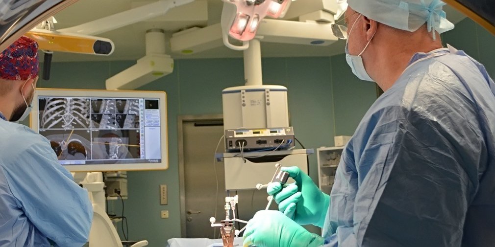
Doctors of Spinal Neurosurgical Centre at Moscow’s Vorokhobov Hospital performed a minimally invasive surgery to remove vertebral hemangioma with a unique technique applied involving high-precision equipment, that is a diagnostic appliance consisting of a mobile CT scanner integrated with the navigation station, Moscow Mayor’s web page reported.
Vertebral hemangioma is a benign vascular tumour affecting bone tissue of the vertebrae. It is removed through percutaneous vertebroplasty, a minimally invasive surgery, when bone cement is injected through the skin into the hemangioma cavity with a thick needle. For that, an X-ray machine is used, but due to the complex spine anatomy, some important structures are almost impossible to visualize with a conventional X-ray machine. In addition, fluoroscopy monitors every surgeon’s move, so both the patient and the surgeon team are exposed to radiation for a long time.
Doctors of Vorokhobov Hospital have used the latest navigation station to perform this procedure. Sensors installed in the surgery room simulated high-precision interactive model of the affected spine section. Every neurosurgeon’s move was displayed on the monitor. Thanks to this, the needle was successfully inserted into a 3.5 mm hole of the narrow spinal canal. The surgery’s success was confirmed by a check CT scanning. The patient had a minimum radiation exposure.
The surgery was performed by Dmitry Dzukayev, Head of Spinal Neurosurgery Centre. He noted that the unique technology was applied thanks to the cooperation between the medical institution and the University Clinic of the Lomonosov Moscow State University.